| ASSAY | RESULT | NOTES |
|
Growth on Tomato Juice Agar (1) |
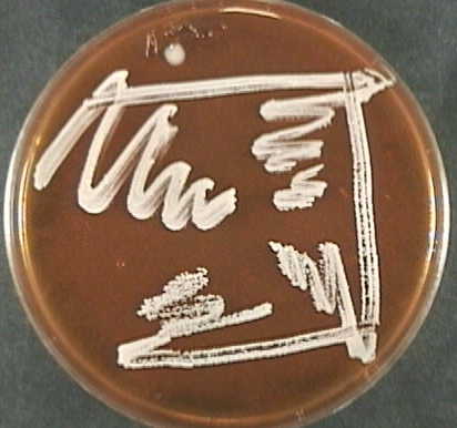 incubated at 37 oC for 48 hours. |
Colony Morphology:
Shape:
|
|
Growth on Tomato Juice Agar (2) |
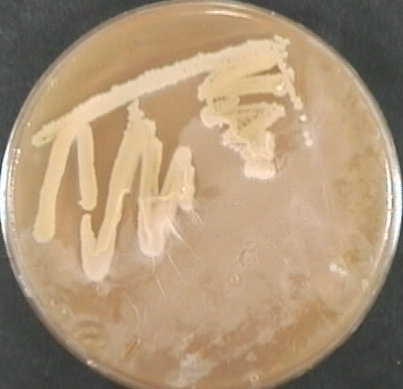 incubated at 37 oC for 48 hours. |
Colony Morphology:
Shape:
|
|
Growth on T-Soy Agar |
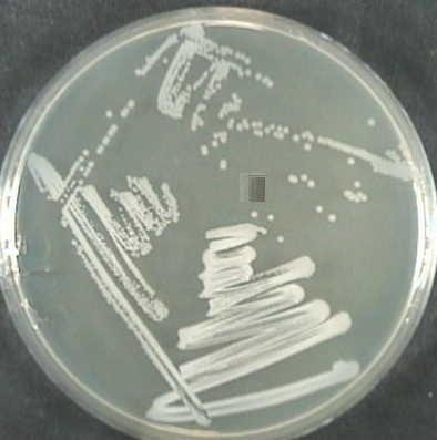 37o C for 48 hours. |
Colony Morphology:
Shape:
|
|
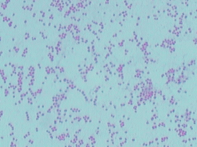
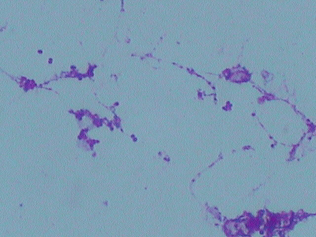
Gram Stain |
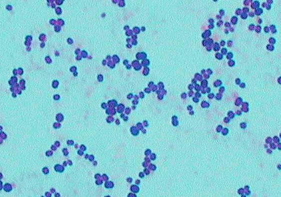 agar. (1000x magnification) |
Gram positive cocci observed |
|
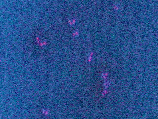
Endospore Stain |
 T-soy agar. (1000x magnification) |
No sporulation observed. Cells stained with safranin. |
|
Capsule Stain |
 agar. (1000x magnification) |
No capsules observed. |
|
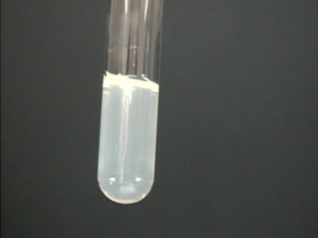
MacConkey's Agar Growth |
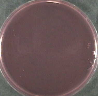 at 37 o C for 48 hours. |
No growth observed. |
|
Sheep's Blood Agar Growth |
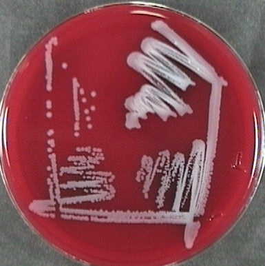 incubated at 37 oC for 48 hours. |
Growth observed, gamma-hemolysis pattern. |
|
Anaerobic Chamber Growth |
 at 37 o C for 48 hours in an anaerobic chamber. |
Colony Morphology:
Shape:
|
| Oxidase Assay | No color change on swab. | Oxidase negative. |
| Catalase Assay | Slight bubble formation. | Catalase negative. |
|
Carbohydrate Utilization Tests |
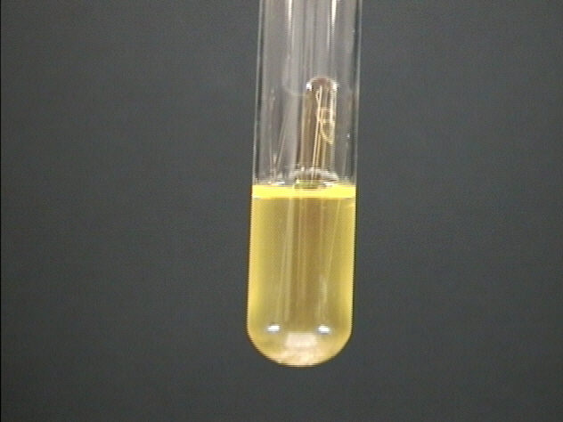 37 o C for 48 hours. 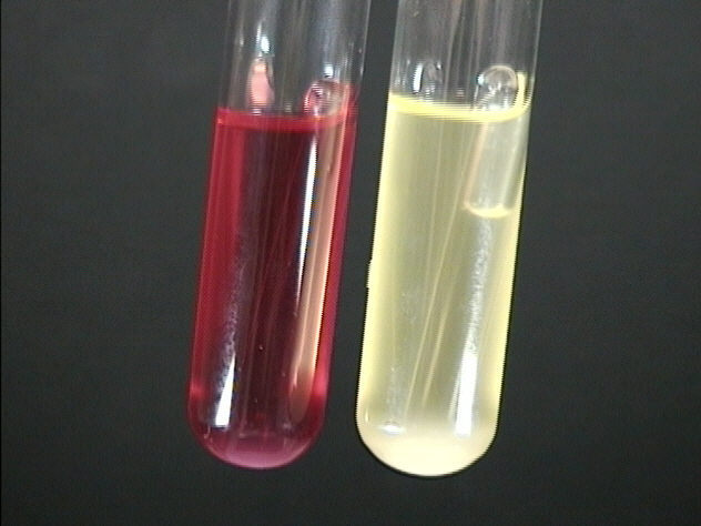 37 o C for 48 hours. Environmental isolate on left . E. Coli control on right. |
Color change and bubble in Durham tube observed (Figure 11).
No color change or bubbles observed in lactose tube innoculated with environmental isolate. Color change and bubble formation in E. Coli control tube. (Figure 12) |
|
Motility Stab |
 at 37 o C for 48 hours. |
Growth confined to stab line. |
| Growth in 4oC | No growth observed. | No growth observed. |
| Growth in 55oC | No growth observed. | No growth observed. |
|
Growth in T-Soy Broth |
 extracted from T-soy broth. (1000x magnification) |
Cellular Arrangement:
|
