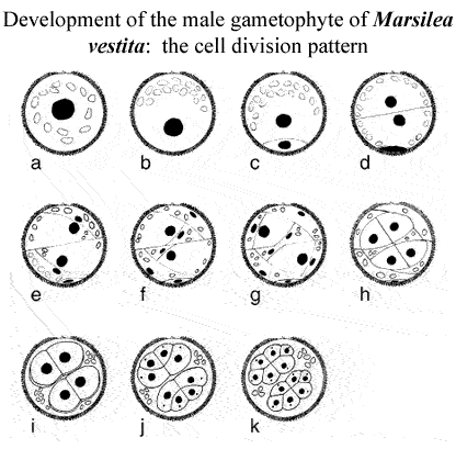
Drawings
of the cell division patterns during the development of the male gametophyte
of M. vestita. (a) Microspores at the time of imbibition. The
nucleus is depicted as a black circle, and the plastids are depicted as
open ellipses. (b) The gametophyte before the 1st division, when cytoplasmic
reorganization occurs. (c) After the 1st mitotic division, a prothallial
cell (bottom) is cut off from the rest of the gametophyte and no longer
divides. (d) The 2nd division gives rise to 2 antheridial initials, essentially
of equal size. (e-g) Each antheridial initial undergoes a series of unequal
division that produce the 3 sterile jacket cells. None of the jacket cells
can proliferate further. (h) Each primary spermatogenous cell undergoes
a symmetric division. (i) The 2 spermatogenous cells in each antheridium
undergo a symmetric division to produce 4 spermatocyte mother cells. By
this stage, the jacket cells become far less conspicuous and they eventually
degenerate. (j) The 4 spermatocyte mother cells in each antheridium undergo
a division to produce 8 spermatocytes. At this stage, a blepharoplast
(dot) appears in each spermatocyte, and then it rapidly disappears (see
Hepler [1976]). Later, the blepharoplasts reform, split and function as
the centrosomes for the next mitotic cycle, when each spermatocyte undergoes
a division that produces 16 spermatids in each antheridium. |