
Textbook Chapter 4
CD Modules NervousSystem I & II

Neuron Structure
Be sure to examine Figures 6.1, 2, 4, &
5. and Table 6.1
It is critical to understand the concepts
behind the terms in this table.
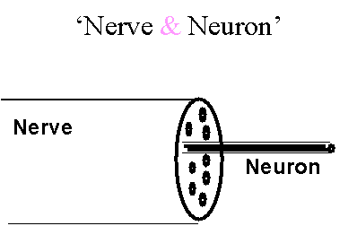
‘Nerve vs. Neuron’
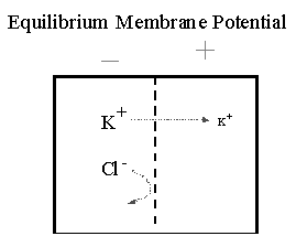
Equilibrium Membrane Potential
(Vm) Made Easy
Membrane Potential (Vm)
See Figs 6.6 - 6.8
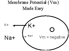
To Approximate Vm:
NERNST Equation
E Ion = R T ln [Ion]1
z F [Ion]2
R = gas constant = 1.987 cal/mol-deg
T = degrees K
z = ion valence
F = Faraday’s Constant 23,062 cal/V-mol
Constants in the Nernst Equation
Temperature
R
F
Log10 = (ln/2.303)
At 37 o C,
EIon (in mV) = - 61.5
log10 [Ion]1
z [Ion]2
At 20 o C,
EIon (in mV) = - 58
log10 [Ion]1
z [Ion]2
What does EIon mean?
Equilibrium Potential (Vm)
Predicting the sign of Vm:
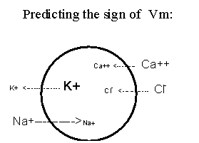
When does Eion approximate Vm?
Use the permeable ion in the Nernst
If it doesn’t diffuse across the membrane,
it doesn’t contribute to Vm
Ohm’s Law
V = I R
I = V/R
When ions (charges) move through membrane
channels, current flows!
Amperes (A)
The flow of ions (I) is impeded by R
We use 1/R or conductance (g) to express
the permeability of a membrane to an ion
Questions:
If GNa+ increases, Vm will……?
If GK+ decreases, Vm will…….?
If GNa+ decreases, Vm will……?
If GK+ increases, Vm will…….?
If [K+]out increases, Vm will…..?
If [K+]in increases, Vm will…..?
If [K+]out decreases, Vm will…..?
If [Na+]out increases, Vm will…..?
If [Na+]out decreases, Vm will…..?
To Calculate Vm: Goldman Equation Text, page 182
Membrane Potential: Changes in Vm:
• Depolarization: change in resting
Vm to a more positive value
• Hyperpolarization: change in resting
Vm to a more negative value

Membrane Potential (Fig 6.9)
Review Graded Potential Fig
6.10
• Variable amplitude
• Variable duration
• Can be summed
• Hyper- or depolarization
• Decremtnatl; not propagated; decreased
amplitude as it passes
along a membrane
Excitable Cells
Conduct action potentials along entire cell
membrane
In humans, nerves, skeletal and cardiac muscle
cells
All possess voltage-gated ion channels
Channels that change between open/closed
states as Vm changes
Voltage-gated ion channels:
Open <---> Closed states determined by
voltage difference across membrane (Vm)
Threshold: Closed state often referred
to as channel inactivation
Has a voltage sensor region
Inactivation region
For Voltage - gated ion channel, see Fig
6.14
Action Potential
(Fig 6.13; Fig 6.15)
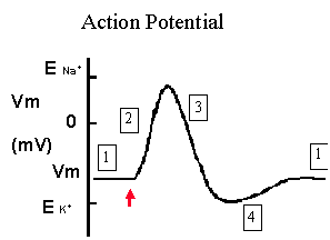
Most nerve cells have voltage gated Na+ channels
At resting Vm, these channels are closed,
Vm is close to E of K+
Voltage gated Na+ channels open when Vm depolarizes
to threshold
Action Potential
• The G of Na+ increases many
fold and Vm goes towards E of Na+
(usually around +30 to +60 mV)
• Na+ channels close and inactivate
• Voltage gated K+ channels open
• Vm returns towards E of K+
Action Potential
• All or None (threshold)
• Same amplitude on a given neuron
• Cannot be summed
• One direction (dendrites to terminal)
• Refractory period
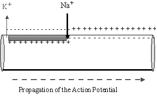
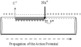
• Propagated (non-decremental)
How does the action potential move along the axon?
How is a depolarization generated at the dendrites
of a neuron?
• Neurotransmitter binding to receptor which
is linked to an ion channel
• Mechanical or other stimulus activating
a sensory structure which
leads to opening an ion channel
How to increase speed at which an action potential
travels down a neuron?
• Increase axon diameter
• Larger diameter has less interior resistance
to current flow
• Depolarization to threshold occurs farther
down the axon, so AP moves
faster in a given period of time
How to increase speed at which an action potential
travels down a neuron?
• Myelinate the axon
• Saltatory conduction
Fig 6.20

Neurotransmiiters & Receptors
• Exocytosis of signal molecule
• Receptors
• [NT] + R <------> [NT]*R
• NT removal
• Graded Potential
• epsp & ipsp
Post Synaptic Events:
EPSP: EXCITATORY POSTSYNAPTIC POTENTIAL
EPSP --> DEPOLARIZATION -->
THRESHOLD -->
ACTION POTENTIAL -->
EXCITATION OF
POST SYNAPTIC STRUCTURE
INHIBITORY - IPSP
IPSP -->
HYPERPOLARIZATION -->
INHIBITION