Ģ Cortex
Ģ Medulla
Ģ Renal Pelvis
Fig 17.2
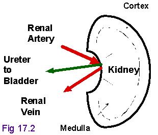
Urinary System: Fig 17.1
Kidney Layers
Ģ Cortex
Ģ Medulla
Ģ Renal Pelvis
Fig 17.2
NEPHRONS
CORTICAL (80%)
JUXTAMEDULLARY (20%)
Vascular component: Blood supply
Tubular:
Filtrate -----> Urine
Fig 17.4
Fig 17.7: Simplified Nephron
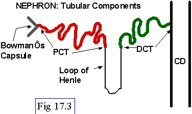
Nephron Components: Tubular
Ģ Glomerulus & BowmanÆs Capsule
Ģ Visceral and parietal layers of B.C.
Ģ Proximal convoluted tubule (PCT)
Ģ Loop of Henle
Ģ Distal convoluted tubule (DCT)
Ģ Collecting Duct
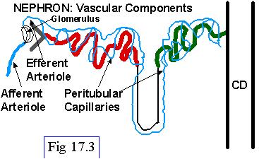
Nephron Components: Vascular
Ģ Afferent and efferent arterioles
Ģ Peritubular capillaries
Ģ Vasa recta
Cortical vs. Juxtamedullary Nephrons
Note Locations in kidney
Peritubular vs. Vasa Recta capillaries
Fig 17.3: The Nephron:
Tubular Component
Fig 17.4: Nephrons:
Cortical & Juxtamedullary
Fig 17.6: Blood Supply to Kidney
Fig 17.6: Vascular Component of Nephron
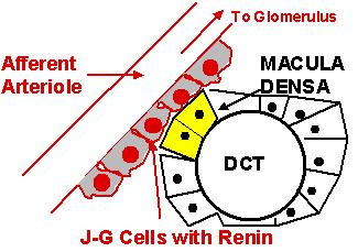
Fig 17.5: Juxtaglomerular Apparatus
Tubular Component: Macula Densa
Ģ Part of DCT
Ģ Juxtaglomerular cells
Ģ Cells of Afferent Arteriole
Ģ Release renin in response to low renal BF and low tubular fluid [Na+]
or [Cl-] content
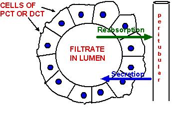
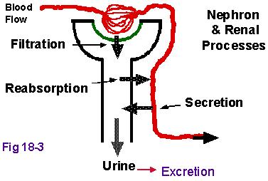
Renal Processes
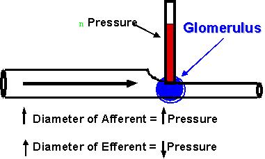
Ģ Filtration
driven by hydrostatic pressure across a selectively permeable capillary
Ģ Reabsorption
solutes moved from filtrate back into blood
Ģ Secretion
solutes moved from blood into filtrate
filtrate becomes urine
Fig 17.7: Renal Processes
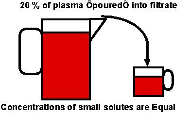
Filtration
Fluid forced from plasma
Approx. 20% of plasma becomes filtrate
Filtrate becomes urine after modifications
Includes solutes small enough to pass through filter-membrane,
i.e., smaller than 69,000 MW
Reabsorption
Desireable substances in filtrate removed and returned to plasma
Active process
Secretion
Remaining undesirable substances in plasma transported into filtrate
Active process