Results


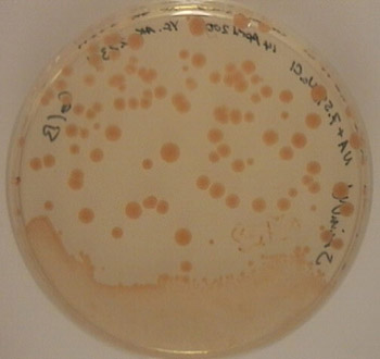
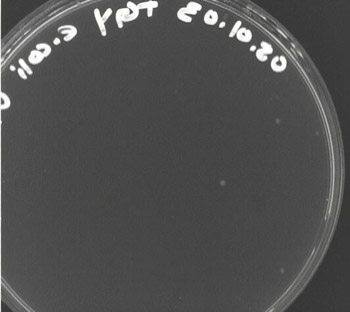
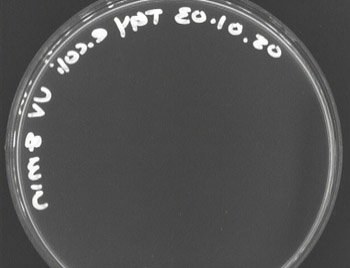
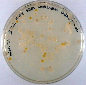
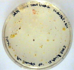
Fig.1  Air Sample Grown on NA + 7.5% NaCl.  It was exposed to
UV for one minute and incubated at 30° C for 2 days and room temperature
for 2 days.  The red colony at the top of the plate was picked for more tests.


gram positive
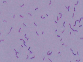
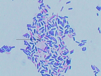
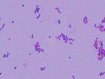
Fig.2  Gram Stain of isolated UV resistant organism
from air sample grown on NA + 7.5% NaCl plate incubated at 30° C
for 5 days.  Shows small gram positive rods.  (1000X)


Fig. 3  A spread plate of the red colony exposed to 5 minutes UV radiation grown on NA + 7.5% salt plates, incubated at 30° C for 7 days.  Colonies are circular, smooth, convex, orangish and opaque.  Fig. 4  A spread plate of the red colony exposed to 10 minutes UV radiation grown on NA + 7.5% salt plates, incubated at 30 °for 7 days.  Fig. 5   A spread plate of the red colony exposed to 15 minutes UV radiation grown on NA + 7.5% salt plates, incubated ° for 7 days. 


Fig. 6  A TGY plate swabbed with E. coli exposed to UV radiation for 2 minutes , incubated at 37° C for 24 hours.  Very small white, opaque and circular colonies. Fig. 7 A TGY plate swabbed with E. coli exposed to UV radiation for 4 minutes, incubated at 37° C for 24 hours. Fig. 8  A TGY plate swabbed with E. coli exposed to UV radiation for 8 minutes, incubated at 37° C for 24 hours.  No visible growth.

Fig. 9  Bird's eye view of motility stab incubated at 30° C for 4 days.  Red pigment on top is the organism growth.

Fig. 10 This is an Gram stain of the red pigmented colony from the air sample.  It was taken from a colony incubated for 7 days.  As the arrow points out, a clear portion inside a Gram positive rod indicates a non-bulging, central endospore.  Previous Gram stains of this organism did not show signs of spores but they were from younger colonies and rods were not as densely clumped.  (1000X)
Fig. 11 This is a spread plate of TGY broth inoculated with desert soil.  The NA + 7.5% NaCl plate was exposed to UV radiation for 15 minutes.  Deinococcus radiodurans was found on this plate after seven days of incubation at 30° C.
Fig.12  This is a Gram stain of a red colony found on the plate in Fig. 11.  The sample came from solid media so we cannot say if the cocci occur in pairs and tetrads.  (1000X)

 Home   Intro     Methods    Discussion
Final Protocol   Acknowledgements   References