STRUCTURE

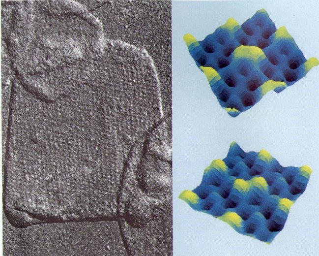
Two-dimensional crystals of VDAC channels in outer mitochondrial membranes
of N. crassa. The left panel is an electron micrograph of
freeze-dried and shadowed outer membranes after treatment with phospholipase
A2 to induce large crystalline arrays. The
crystalline pattern is visible on the surface of the central flattened
vesicle with straight edges. On the right are the results of computer
filtration, everaging and image reconstruction. The false color
indicates elevation (bright yellow being high and dark blue being low).
The two images represent the two surfaces of the crystal. Each image
contains four six-channel repeating units. The figure is adapted
from the work of Thomas et al. The color images were produced
by B.L. Trus.

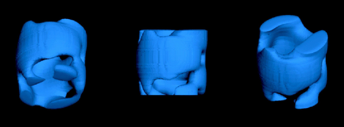
This reconstructed image of the structure of the wall of N. crassa VDAC is courtesy of Carmen Mannella:
C.A. Mannella (1997) J. Bioenerg. Biomembr. 29: 525-531.
It shows where negative stain protrudes beyond the central cylinder.
Please
send comments and contributions to Dr.
Marco Colombini |
Last modified: April 23, 2004 12:50 Page created and maintained by Swapnil Sharma |